FERTILITY PROFILE - Hormones / Cortisol Function / Thyroid hormones / Women's Health / Fertility
Indications:
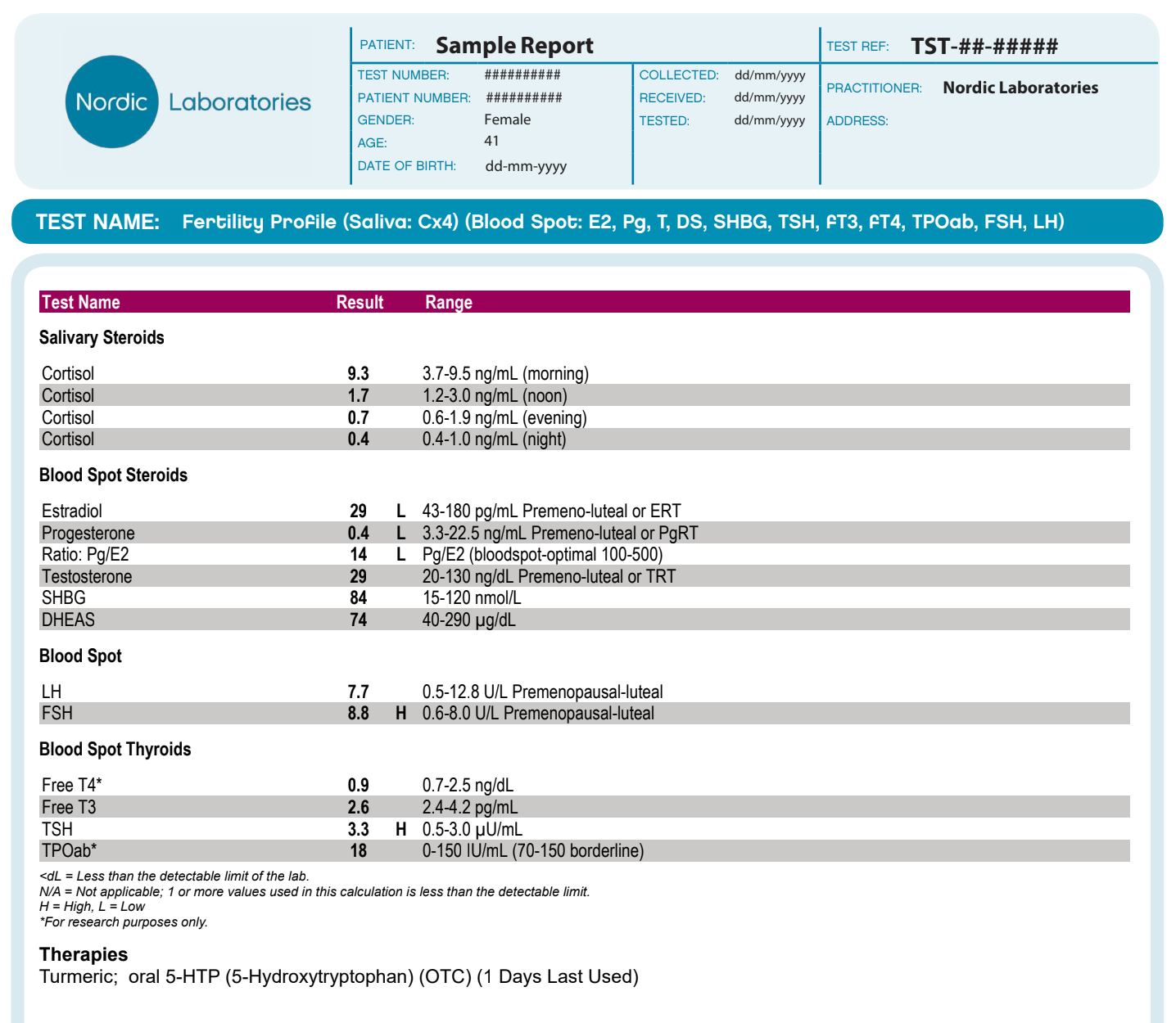
The Fertility Profile is a bloodspot and saliva test that measures hormone levels that contribute to female fertility.
In North America, many couples are having difficulty getting pregnant. Current statistics from the most recent National Survey of Family Growth show that 7.4% (2.1 million) of all married women of childbearing age (15-44 years) in North America are infertile, defined as not having achieved pregnancy despite using no contraception in the past 12 months or more. When the survey considered only married, childless women, this figure increases to 16.6%. The incidence of female infertility is significantly age-related, increasing with age. In one population study, the main causes of infertility attributable to the woman were ovulatory failure (21%), tubal damage (14%), and endometriosis (6%), while a massive 28% of cases were unexplained. Yet in the absence of a physical cause, many cases of female infertility may be explained by something as simple as a correctable hormone imbalance, which can be assessed by hormone testing.
Overview
Analytes measured:
- Estradiol (E2)
- Progesterone (Pg)
- Testosterone (T)
- DHEA-S
- Diurnal cortisol (C) sampled four times during a day
- Thyroid hormones (free T3, free T4, TSH, and TPO antibodies)
- LH
- FSH
Profile II tests all of the hormones in bloodspot with the exception of the 4x diurnal C tested in saliva. Sampling is done on days 19-21 of the menstrual cycle, coinciding with the mid-luteal phase, when the level of progesterone should be optimal for a successful pregnancy.
Hormonal Aspects of Infertility:
Hormone-related causes of female infertility most often involve the following five scenarios:
- Ovarian Insufficiency
While the average age of menopause is 51, women can start experiencing signs of ovarian insufficiency even in their thirties. A cessation of ovulation prior to the age of 40 is rare, and is usually referred to as premature ovarian failure. Declining ovarian function is the main reason for the age-related decline in female fertility. As the number of available follicles starts to fall, estrogen is still being produced but ovulation does not occur, and progesterone levels fall in the absence of a corpus luteum. Also, while high FSH levels on day 3 of the menstrual cycle typically confirm premature ovarian failure and the onset of menopause, FSH levels remain elevated on day 19-21 compared to those seen in normal ovulatory cycles, and despite normal LH values. A typical pattern of day 19-21 hormone levels indicating signs of ovarian insufficiency would consist of: low estradiol, low progesterone, low testosterone and elevated FSH; LH, DHEA-S, cortisol and thyroid hormones may or may not be normal.
- Luteal Phase Deficiency
In some patients who are infertile, ovulation may occur normally but levels of progesterone are inadequate during the luteal phase. This luteal progesterone deficiency means that, even if the egg is fertilized, implantation either does not occur, or if it does the progesterone produced by the corpus luteum is not high enough to sustain the pregnancy. Luteal phase deficiency can be caused by a number of problems, including endometriosis and abnormal follicular development, but most commonly it is a result of inadequate progesterone production by the corpus luteum. A typical finding is low progesterone levels in the luteal phase, usually with normal estradiol levels.
- Polycystic Ovarian Syndrome (PCOS)
PCOS is the most common endocrine disorder affecting women of reproductive age, and is closely associated with insulin resistance, metabolic syndrome, and future risk of developing diabetes and cardiovascular disease. Among women presenting with infertility in one study, PCOS was found to be present in 81% of women who were anovulatory, in 50% of those with tubal disease, and in 44% of those with unexplained infertility. PCOS is diagnosed when a patient has two of these three criteria: polycystic ovaries on ultrasound; oligo- and/or anovulation; and hyperandrogenism9. Hormonally, it is characterized by low progesterone, normal-to-high estradiol, high testosterone, normal to high DHEA-S, and LH elevated 2-3 times relative to FSH. Cortisol and thyroid hormones may or may not be normal, although women with PCOS have been found to have a three-fold higher prevalence of autoimmune thyroiditis compared to healthy women.
- Hypometabolism/Thyroid Deficiency
Thyroid dysfunction, including subclinical hypothyroidism (elevated TSH with normal free T3 and free T4 levels), has been implicated as a cause of infertility, yet thyroid hormone treatment can be a simple solution to restore a regular menstrual pattern. In one study, levothyroxine treatment resulted in pregnancy in 44% of infertile patients diagnosed with subclinical hypothyroidism. In patients with hypometabolism as a result of thyroid dysfunction, the sex hormones, LH and FSH and cortisol may all be normal but the patient has symptoms of hypothyroidism, and a low T3 level. High TPO antibodies indicate an autoimmune thyroid disease (e.g., Hashimoto’s Disease), which is associated with fertility-related problems, and it is important to rule out thyroid autoimmunity in women attempting to conceive because of the increased risk of miscarriage.
- Stress
Stress can severely affect a woman’s ability to conceive, probably because of its impact on endocrine balance, particularly affecting thyroid and adrenal function and the pituitary’s production of gonadotrophs. The diurnal cortisol variation measured in saliva samples collected on waking, late morning, late afternoon and at bedtime, indicate the effects of stress on adrenal function as well as the adrenal glands’ ability to produce cortisol, a hormone important for the cellular actions of thyroid hormones. Stress stimuli also increase prolactin production, and high cortisol and prolactin levels are seen in endometriosis. Endometriosis is found in more than 50% of women with unexplained infertility, and the high cortisol and prolactin levels induced by stress have been implicated in the development of this condition.


